Hemo-flagellate
Including genus: Leishmania and Trypanosoma(blood tissue species):
There are four morphological forms of clinical significance associated with the hemoflagellates:
- Amastigote
- Promastigote
- Epimastigote
- Trypomastigote.
General characteristics
- They live in the blood and tissues of man and other vertebrate hosts and in the gut of the insect vector.
- Members of this family have a single nucleus, a kinetoplast and a single flagellum.
- Nucleus is round or oval and is situated in the central part of the body.
- Kineloplast consists of a deeply staining para-basal body and adjacent dot-Like blepharoplast.
- The parabasal body and blepharoplast are connected by one or more thin fiber.
- Flagellum is a thin, hair-like structure, which originate from the blepharoplast.
- The portion of the flagellum ,which is inside the body of the parasite and extends from the blepharoplasl to surface of the body is known as axoneme.
- A free flagellum at the anterior end traverses on the surface of the parasite as a narrow undulating membrane.
- Hemoflagellates exist in two or more of four morphological stages.
The transmission of hemo-flagellates
Is accomplished by the bite of an arthropod vector. Flagellate protozoa found in blood or tissues of human and there are two genera of medical importance (Leishmania and Trypanosoma).
The major difference between these two genera is that primary diagnostic form found in Leishmania is the amastigote, whereas that of Trypanosoma is the trypomastigote.
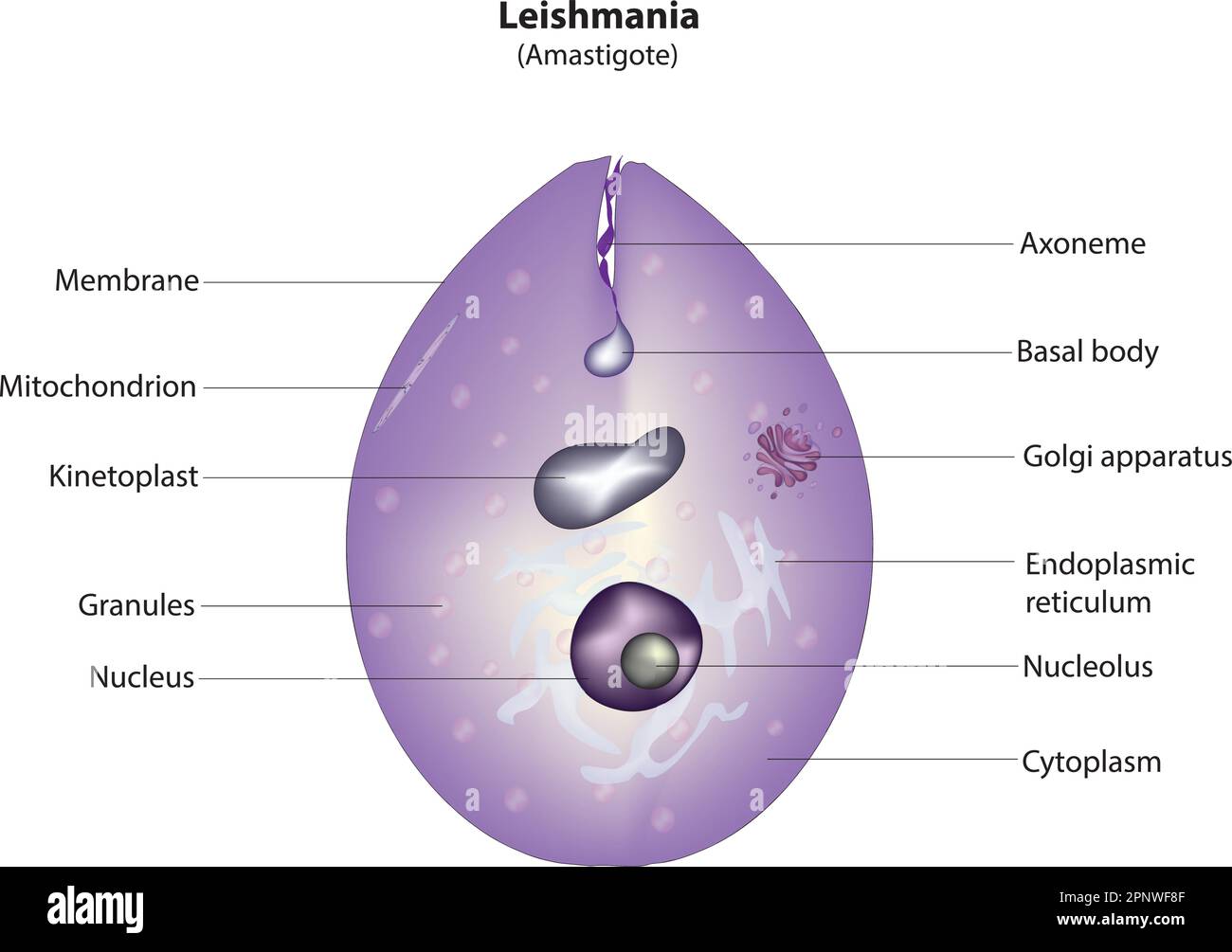
- Amastigotes:
- It is Roundish to oval in shape, Consist of a nucleus and kinetoplast.
- The large single nucleus is typically located off-center .
- The dot-like blepharoplast is attached to a small axoneme, this axoneme extends to the edge of the organism.
- The single parabasal body is located adjacent to the blepharoplast.
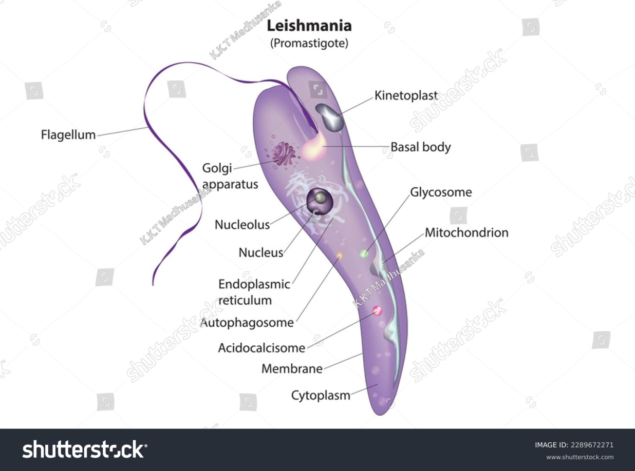
- Pro-mastigotes:
- It is Long and slender in appearance.
- The large single nucleus is located in or near the center .
- The kinetoplast is located in the anterior end of the organism .
- A single free flagellum extends anteriorly from the axoneme.
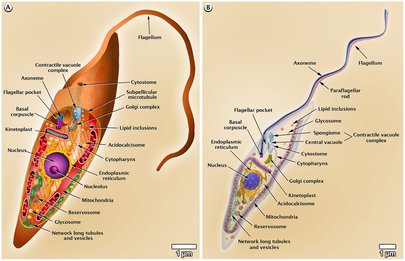
- Epimastigotes:
- It is long and slightly wider than promastigote form.
- The large single nucleus is located in posterior end .
- The kinetoplast located anterior to the nucleus .
- Undulating membrane extending half of the body length .
- A single free flagellum extends anteriorly from the axoneme.
- Trypomastigotes:
- It is C or U shape in stained blood films .
- Long and slender in appearance .
- One nucleus located anterior to the kinetoplast .
- The kinetoplast is located in the posterior end of the organism .
- Undulating membrane extending entire body length .
- A single free flagellum extends anteriorly from the axoneme when present.
Genus Leishmania:
Leishmaniasis
There are many different species of Leishmania and the disease that they cause. directly linked to the species of Leishmania with which a person Several species of Leishmania are pathogenic for man: L. donovani causes visceral leishmaniasis (Kala-azar, black disease, dumdum fever); L. tropica cause cutaneous leishmaniasis (oriental sore, Delhi ulcer, Aleppo, Delhi or Baghdad boil); and L. braziliensis (also, L. mexicana and L. peruviana) are etiologic agents of mucocutaneous leishmaniasis .
Life Cycle of Leishmania sp.
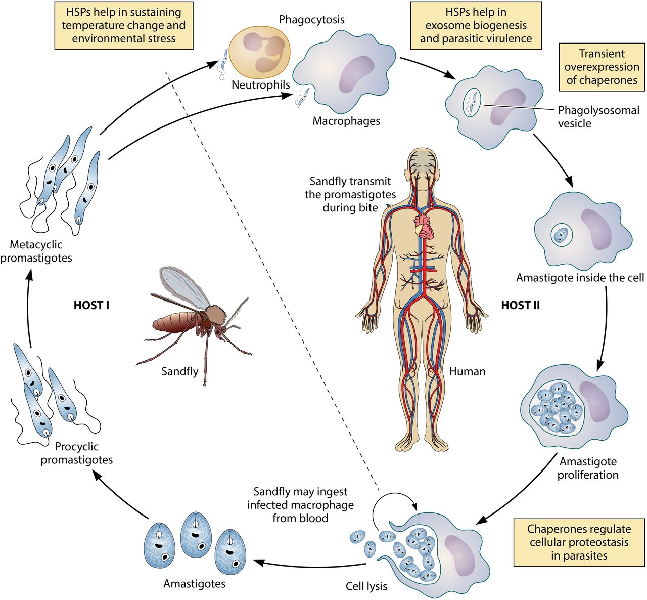
- Leishmania are transmitted by arthropod.
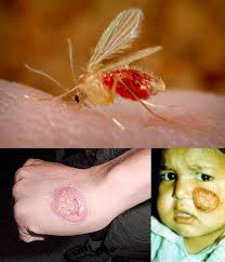
- In this case it is a small biting fly known as a sand fly.
- Leishmaniae spend part of their life cycle in the gut of the sandfly, but their life cycle is completed in a vertebrate host.
- Within the sandfly gut, the protozoa are carried as extracellular pro-mastigotes , these parasites multiply in the gut and migrate toward the pharynx.
- Sandflies transferred pro-mastigotes to the vertrebrate host when the sandfly takes a meal blood by expelling Leishmania into the bite wound of the mammalian host.
- From where they pass into the blood and tissues of the human host.
Pathogenesis and clinical finding
Leishmaniasis is a parasitic disease caused by several species of genus
Different species of leishmania cause different disease.
- L. donovani causes visceral leishmaniasis also called Kala-azar and Dum Dum fever. Spleeno-megaly and hepatomegaly the infection is generalized and the parasite is distributed in the internal organs. The parasite may also cause a variety of skin lesions (dermal leishmaniasis) without any visceral manifestations.
Laboratory Diagnosis
1. Giemsa-stained slides of blood , bone marrow , lymph node aspirates and biopsies of the infected areas for the diagnosis of amastigote forms.
2. Culture of blood, bone marrow and other tissues these samples show the promastigote forms.
3. Serological tests ( Detect of Antibodies )
B. L. tropica: causes tropic sore or Baghdad boil, oriental sore and cutenaeous Leishmianiasis.
The infection is limited to a local lesion of the skin and subcutaneous tissues.
Laboratory Diagnosis
1. The specimen of choice for identify the amastigotes of Leishmania braziliensis is a biopsy of the infected ulcer.
2. Microscopic examination of the Giemsa-stained preparations should reveal the typical amastigotes. Promastigotes may be present when the sample is collected immediately after introduction into the patient.
3. Culturing the infected material, which often demonstrated the promastigote stage.
4. Serological tests.
Treatment
Pharmacologic therapies include the following:
· Pentavalent antimony (sodium stibogluconate or meglumine antimonate): Used in cutaneous leishmaniasis
· Liposomal amphotericin B (AmBisome): Effective against pentavalent antimony ̶resistant mucocutaneous disease and visceral leishmaniasis .
American trypanosomes
1-Trypanosoma cruzi
1-life cycle: in direct life cycle
2-final host: man
3-Resevore host: dogs, cat and rat
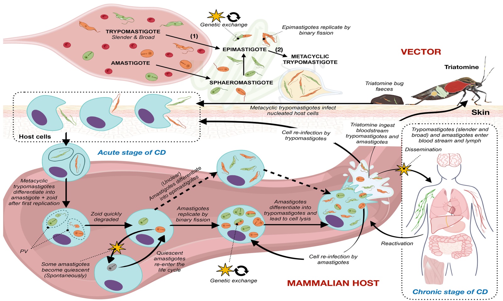
4-Intermediat host : Reduviid bug
5-infective stage :Metacyclic form
6-Dignostic stage: Trypomastigote
7-Disease name: Chagas disease
Note: this parasite contains all stages of Trypansoma : Amastigote, Promastigote, Epimastigote, Trypomastigote, Metacyclic form.
8- Habitat: blood, Csf, and lymph
Pathogenesis of Trypanosoma cruzi
Stages of the disease
1-Acute stage: the presence of pseudo-cyst in every kind of tissue in the body.
Symptoms: anemia ,muscle and bone pain, heart failure and death may occurs with (3-4) in untreated cases and it is most common among children below 5 years.
2-chronic stage: it is most common in adult.
Symptoms: central and peripheral nervous dysfunction with heart enlargement .
Diagnosis of Trypanosoma cruzi
1-Microscobical examination:
A-Demonstrations of trypomastigote in blood film , lymph nodes, bone marrow aspirate.
b-demonstration of amstigote in aspiration of visceral organs.
2-Serological test
3-PCR
4-Xeno-dignosis :experimental examination.
African trypanosomes
1-Trypanosoma gambiense(west African sleeping sickness)
2-Trypanosoma rhodesiense (East African sleeping sickness)
General information
- 1-life cycle: in direct life cycle
- 2-final host: man
- 3-Resevore host: Domestic animals
- 4-Intermediat host : Testse Fly(Glossina)
- 5-infective stage :Metacyclic form
- 6-Dignostic stage: Trypastigote
- 7-Disease name:
1-Trypanosoma gambiense (west African sleeping sickness)
2-Trypanosoma rhodesiense (East African sleeping sickness)
- 8- Habitat: blood, CSF , and lymph
Pathogenesis of African Trypanosoma
Trypanosomes live in blood, lymph nodes, spleen – therefore not intracellular , Particularly abundant in intercellular spaces in brain Humans
Local reaction: painful sore at site of bite, disappears after a couple of weeks.
Trypanosomes reproduce rapidly once enter blood and lymph system – generalized invasion of all organs .
Stages of the disease
1-Parasitemia: the presence of parasite in the blood
2-Lymphadenitis: occur in lymph nods of the base of skill ,legs and show enlargement and congestion and enlargement of liver and spleen.
3- CNS involvement: commonly referred as sleeping sickness during eating or standing or working and finally Death .
Diagnosis
1-Microscobical examination: to see trypomastigote in blood film, lymph nodes , CSF and bone marrow.
2-Serological test
3-PCR ) polymerase chain reaction )

