Nonpathogenic Intestinal Ameba
Entamoeba dispar
- Entamoeba dispar is morphologically indistinguishable (both cyst and
trophozoite) from E. histolytica, so it may be considered as a subspecies of
E. histolytica.
2. It can be distinguished from E. histolytica by: Zymodeme
study (hexokinase iso-enzyme pattern), molecular methods, PCR amplifying, detection of lectin antigen in stool, and RBC inside trophozoites—present only in E. histolytica.
3. E. dispar parasite is nonpathogenic, usually colonizes in the large intestine (10 times more than E. histolytica) but doesn’t invade intestinal mucosa.
4.It grows well in polyxenic media, however, poorly grows on axenic media. E. dispar doesn’t induce antibody production.
Life Cycle and Morphology
The life cycle is essentially identical to that of Entamoeba coli or any of the other nonpathogenic intestinal protozoa, and the cyst form is the infective form for humans.
Morphology of Trophozoites
Living trophozoites vary in diameter from about 12 to 60 μm. Motility has been described as rapid and unidirectional, with pseudopods forming quickly in response to the conditions around the organism; it may appear to be sporadic. Although this characteristic is often described, it is rare to diagnose these organisms on the basis of motility seen in a direct wet mount.
The cytoplasm is differentiated into a clear outer ectoplasm and a more granular inner endoplasm. Based on the recent ability to culture these organisms in axenic culture systems and on light and electron microscopy studies, there may be some morphologic differences between E. histolytica and E. dispar. However, these differences would not be recognized by routine diagnostic methods such as the permanent stained smear. If organisms were seen that were consistent with E. histolytica /E. dispar on the permanent stained smear, the laboratory report would indicate that fact and would be written as “Entamoeba histolytica /E. dispar”
When the organism is examined on a permanent stained smear (trichrome or iron hematoxylin), the morphological characteristics are easily seen.
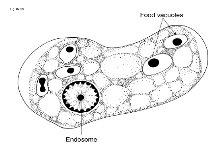
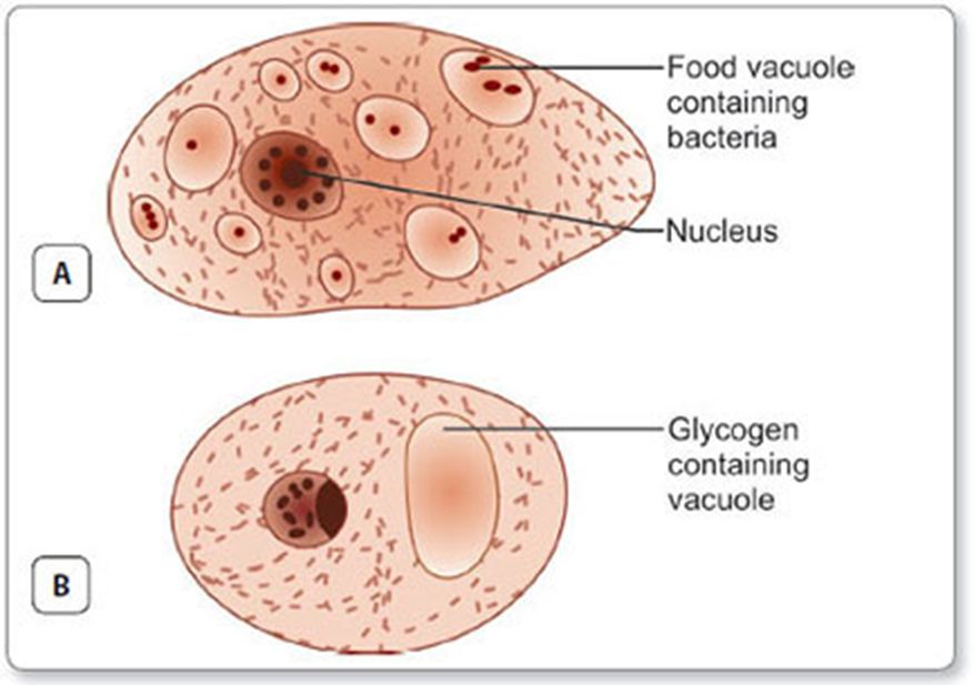
- The nucleus generally has evenly arranged chromatin on the nuclear membrane and has a small, compact, centrally located karyosome.
- The cytoplasm is described as finely granular with few ingested bacteria or debris in vacuoles.
- Ingested RBCs are never seen in the trophozoites; if ingested RBCs are seen, this finding identifies the organism as E. histolytica, not E. dispar.
Entamoeba moshkovskii
Entamoeba moshkovskii is also morphologically indistinguishable from E. histolytica and E. dispar (may be the third subspecies of E. histolytica). This species was first described from Moscow sewage by Tshalaia in 1941 and was thereafter reported to occur in many different countries including India. It can be distinguished from E. histolytica by isoenzyme analysis, molecular methods, and detection of lectin antigen. Though it is a non-pathogen harboring in the intestine recent studies from Bangladesh and India have reported E. moshkovskii as a sole potential pathogen in patients presenting with gastrointestinal symptoms and/or dysentery, highlighting the need for further study to investigate the pathogenic potential of this organism.
Entamoeba moshkovskii is found worldwide and is generally considered to be a free-living ameba. Based on microscopic morphology, this organism is indistinguishable from E. histolytica and E. dispar, except in cases of invasive disease when E. histolytica contains ingested RBCs.
Although first isolated from sewage, E. moshkovskii can also be found in clean riverine sediments to brackish coastal pools. Apparently, there are some differences that separate this organism from E. histolytic and E. dispar. However, these differences pertain to physiology rather than morphology; E. moshkovskii is osmo-tolerant, can be cultured at room temperature, and is resistant to emetine.
Life Cycle and Morphology
The life cycle is essentially identical to that of E. dispar, and morphological differences are minimal to none. In wet preparations, trophozoites usually range in size from 15 to 20 μm and cysts normally range in size from 12 to 15 μm. It is important to remember that on the permanent stained smear there is a certain amount of artificial shrinkage due to dehydration; therefore, all of the organisms, including pathogenic E. histolytica, may be somewhat smaller (from 1 to 1.5 μm) than the sizes quoted for the wet-preparation measurements.
Morphology of Trophozoites
Trophozoites do not ingest RBCs, and the motility is similar to that of both E. histolytica and E. dispar. Nuclear and cytoplasmic characteristics are very similar to those seen in E. histolytica; however, trophozoites of E. moshkovskii do not contain ingested RBCs.
Morphology of Cysts
Nuclear characteristics and chromatoidal bars are similar to those in E. histolytica and E. dispar.
Entamoeba Coli
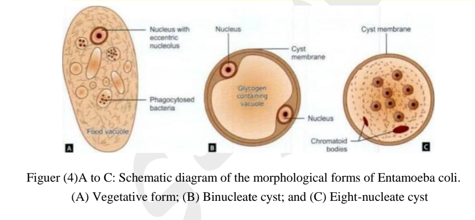
Entamoeba coli was first described by Lewis (1870) and Cunningham (1871) in Kolkata and its presence in healthy persons was reported by Grassi (1878). It is worldwide in distribution and a nonpathogenic commensal intestinal ameba. It is a larger than E. histolytica about 20-50 µm with sluggish motility and contains ingested bacteria but no red cells. The nucleus is clearly visible in unstained films and has a large eccentric karyosome and thick nuclear membrane lined with coarse granules of chromatin.
Cysts are large, 10- 30 µm in size, with a prominent glycogen mass in the early stage. The chromatoid bodies are splinter-Like and irregular. The mature cyst has eight nuclei (8).

Life cycle
The life cycle is the same as in E. histolytica except that it remains a luminal commensal without tissue invasion and is nonpathogenic.
Entamoeba hartmanni
- Entamoeba hartmanni occurs wherever E. histolytica is found.
- It is now considered to be a separate species of nonpathogenic commensalintestinal ameba.
- It is much smaller than E. histolytica, the trophozoirc measuring 4- 12 µm and cyst 5-10 µm in size.
- Trophozoites do not ingest red cells and their motility is less vigorous.
- The cyst resembles that of Endolimax nana.
Entamoeba Gingivalis
- Entamoeb gingivalis was the first ameba of humans
- discovered by Gros in 1849.
- It is global in distribution. Only the trophozoite is found; the cystic stage being apparently absent.
- The trophozoite is about 10-20 µm, actively motile with multiple pseudopodia.
- The cytoplasm contains food vacuoles with ingested bacteria, leukocytes, and epithelial cells.
- Nucleus is round with central karyosome lined by coarse chromatin granules.
- The ameba lives in gingival tissues and is abundant in unhygienic mouths.
- It is a commensal and is not considered to cause any disease.
- It is transmitted by direct oral contact.
- E. gingivalis have been found in bronchial washings and vaginal and cervical smears, where it can be mistaken for E.histolytica.

Endolimax nana
- This common commensal ameba is widely distributed.
- It lives in the human intestine.
- The trophozoite is small (nana: small), less than 10 µm in size with sluggish motility.
- The nucleus has a conspicuous karyosome connected to the nuclear membrane by one or none coarse strands.
- The cyst is small, oval, and quadrinucleate with glycogen mass and chromidial bars, which are inconspicuous or absent.
- It is nonpathogenic.
Iodamoeba Butschlii
- This is widely distributed, though less common than E. coli and E. nana.
- Trophozoite is small, 6- 12 µm, with a conspicuous nucleus.
- The prominent karyosome is half the size of the nucleus, having a bull's eye appearance.
- The cyst is oval, uni-nucleate and has a prominent iodine staining glycogen mass (iodophilic body) Hence, the name lodamoeba.
- It is nonpathogenic.


